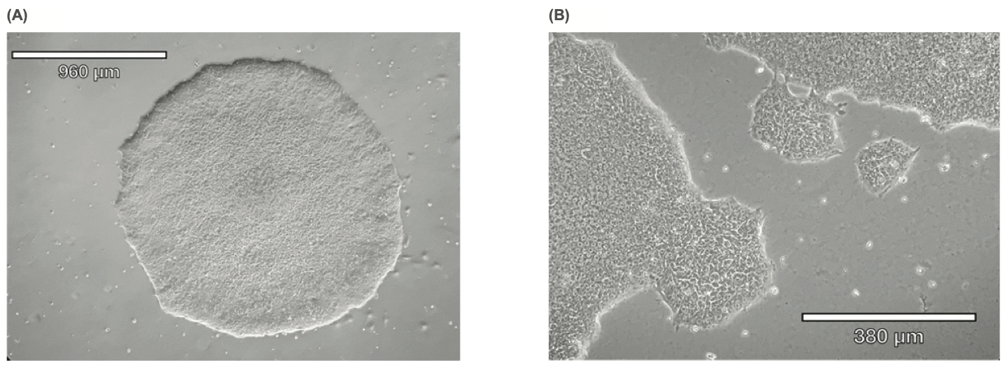
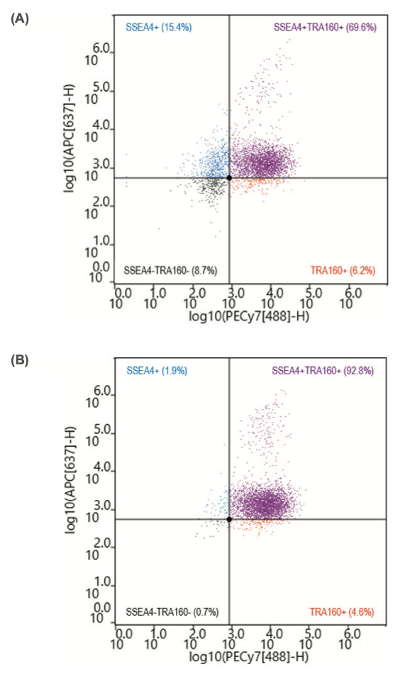
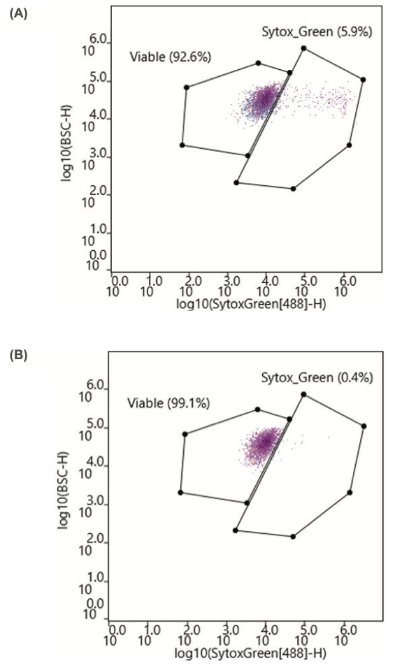
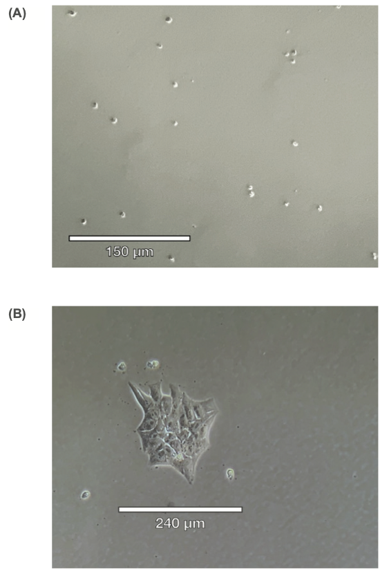
Isolating Induced Pluripotent Stem Cells by Using the WOLF G2 to Sort Stemness-Related Surface Markers

NanoCellect Biomedical, Inc., San Diego, CA
Defined Bioscience, Inc. San Diego, CA
If you're interested in learning more, we’d love to hear from you.

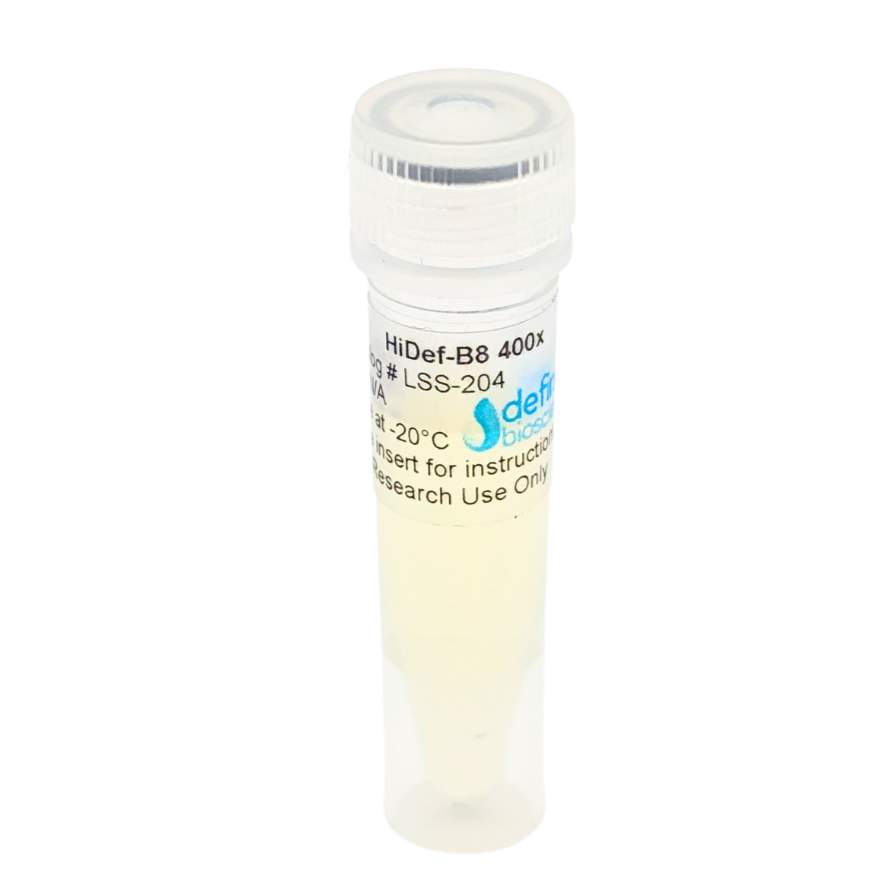

HiDef® B8 Stem Cell Growth Medium
Regular price
From $150.00
Sale price
From $150.00
Regular price
$150.00
Choose your option


DMEM/F12 Basal Medium
Regular price
From $40.00
Sale price
From $40.00
Regular price
$40.00
Choose your option

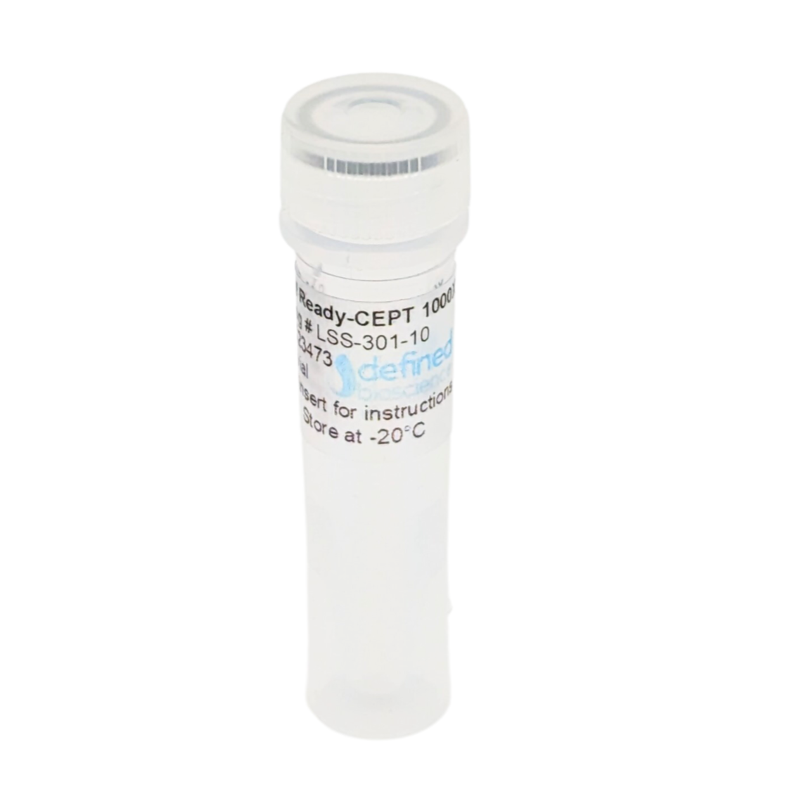
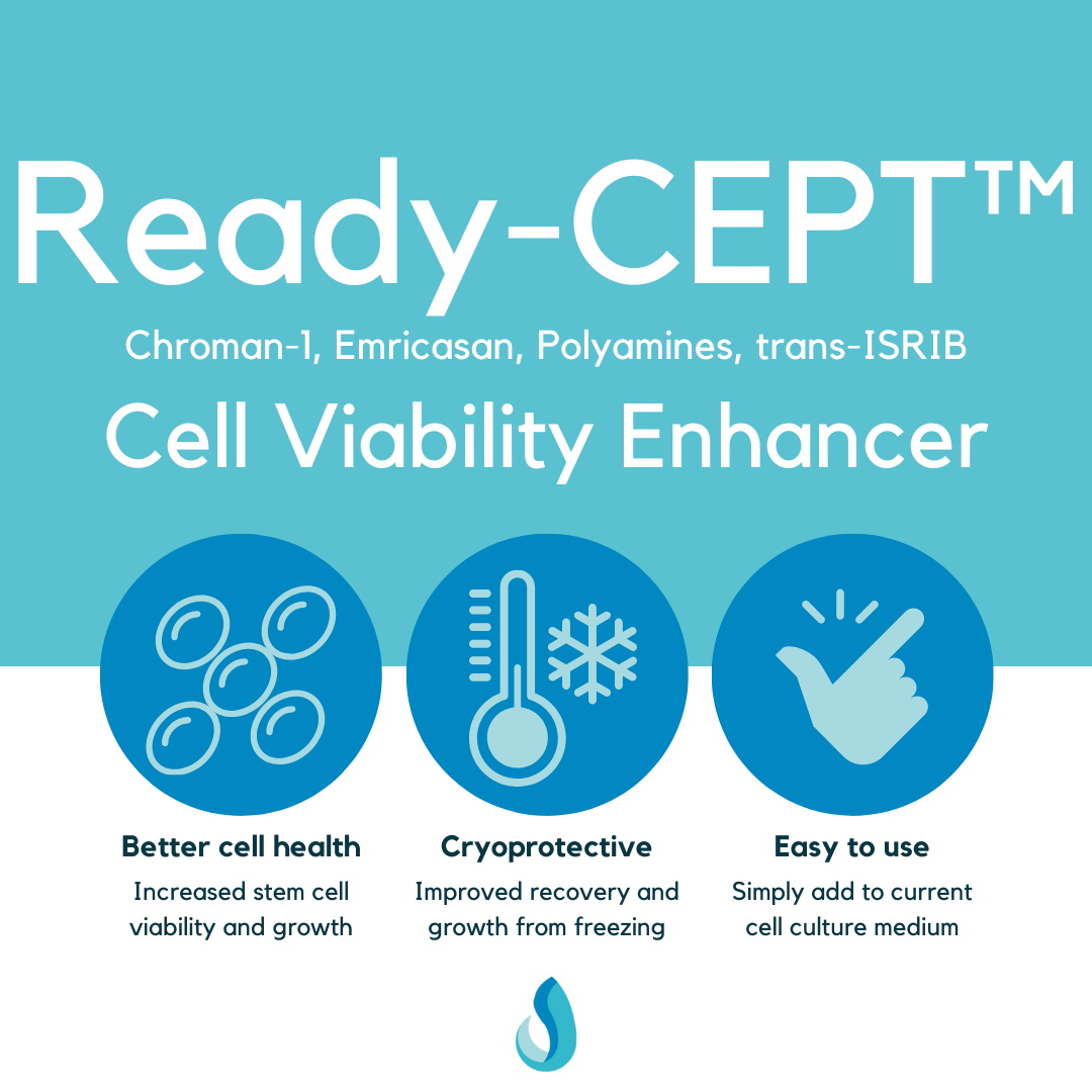
Ready-CEPT® Cell Viability Enhancer
Regular price
From $150.00
Sale price
From $150.00
Regular price
Choose your option

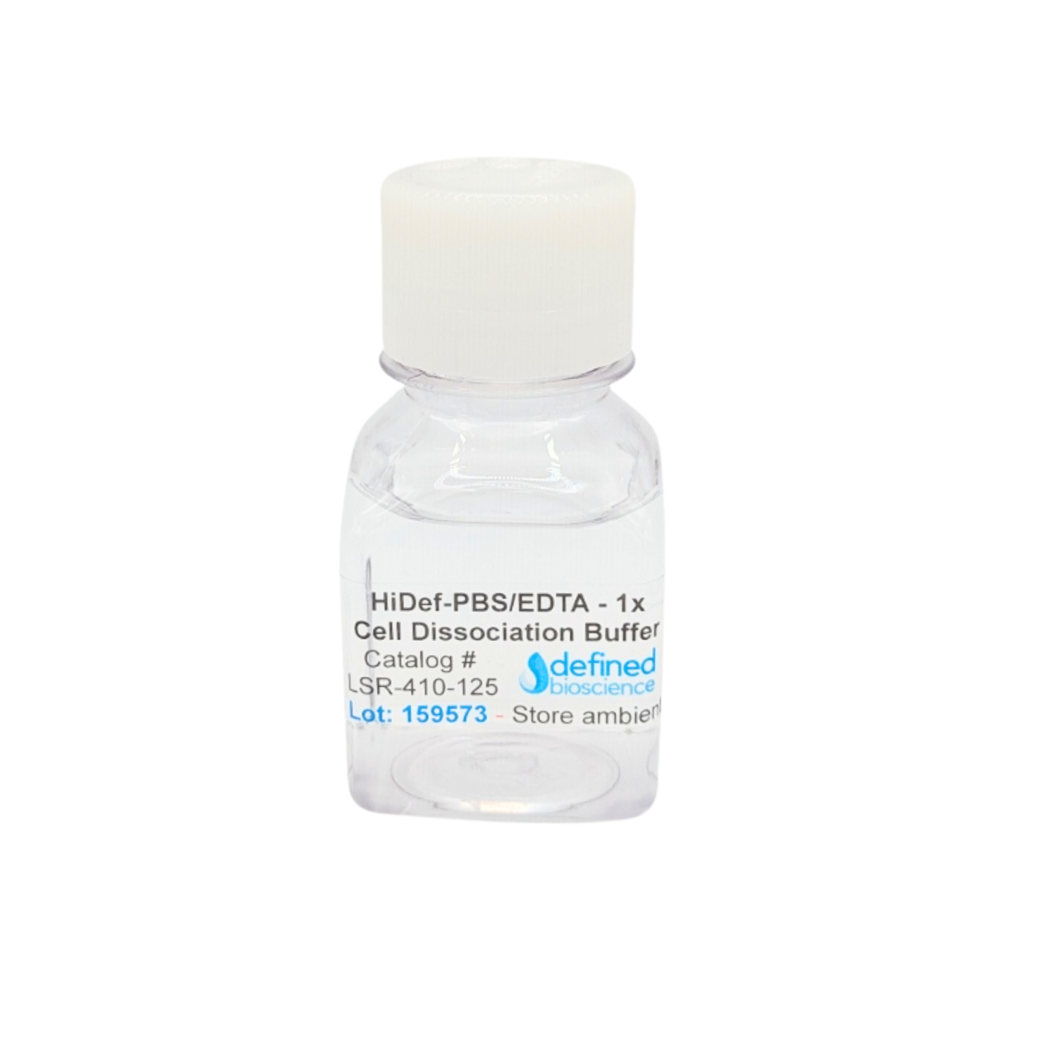
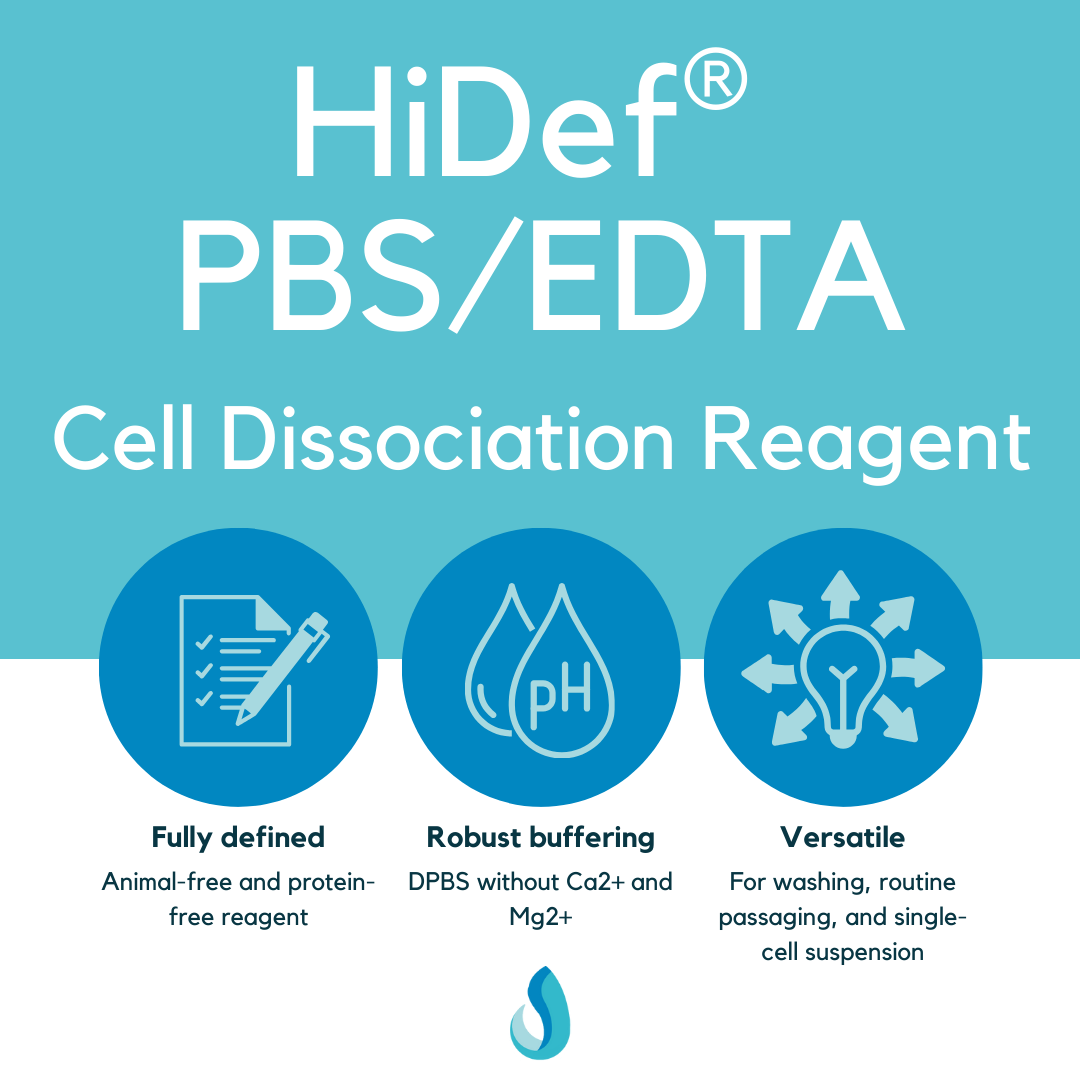
HiDef® PBS/EDTA Cell Dissociation Reagent
Regular price
From $18.00
Sale price
From $18.00
Regular price