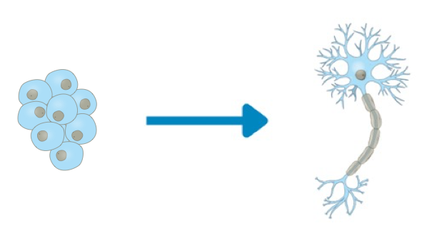
iPSC generation
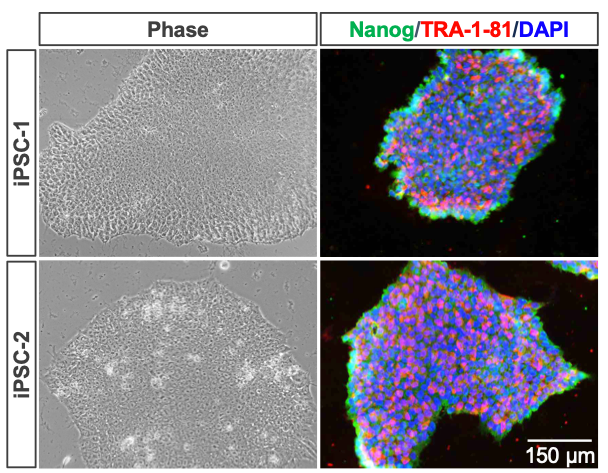
This study leverages iPSCs derived from a female donor ("iPSC-1") and an unrelated male donor ("iPSC-2") to ensure robustness and biological relevance from distinct genetic backgrounds across both sexes. iPSCs were cultured using HiDef-B8 (Defined Bioscience), a serum-free, defined medium optimized to maintain pluripotency and promote robust cell growth. These cells were expanded under feeder-free conditions, single cell-passaged with Accutase™ (Innovative Cell Technologies) or aggregate-passaged with collagenase IV. iPSC lines were verified using key pluripotency markers independent of those used for reprogramming including Nanog and TRA-1-81 (Figure 1). These karyotypically stable iPSCs serve as the foundation for downstream neural differentiation, supporting the development of both 2D and 3D neural differentiation workflows (Figure 2).
Neural Differentiation Workflows
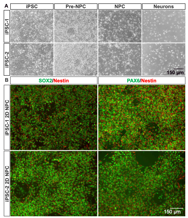
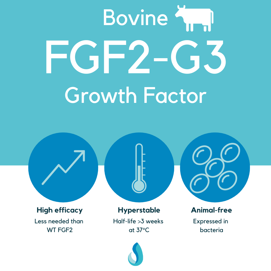
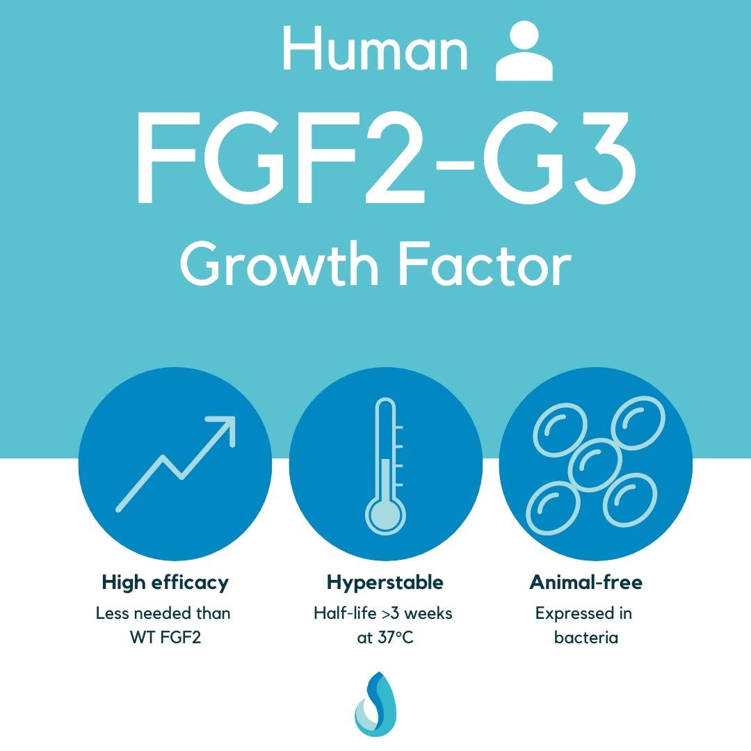
2D Differentiation Workflow (Figure 2, Upper Panel): The 2D workflow, based on Chambers et al. (2009) and updated in Manos et al. (2022), employs a monolayer culture system starting with iPSCs grown to confluency in HiDef-B8 medium and then briefly in HiDef-B6, formulated without FGF2-G3 and TGFβ3. Neural induction is initiated by treating the cells with SMAD inhibitors (SB432542 and LDN193189), promoting a direct and efficient transition to neural progenitor stages. After replating, the cells are expanded as NPCs and then differentiated into neurons using Neurobasal™ (Thermo-Fisher) medium supplemented with B27, alanyl-glutamine, and key neurotrophic factors (BDNF, GDNF, d-cyclic AMP, ascorbic acid, and laminin) and small molecules (RO4929097, cyclopamine, LDN193189). This streamlined, high-throughput- compatible method provides a practical platform for modeling neural processes in vitro.
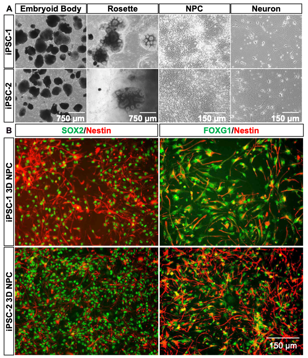
3D Differentiation Workflow (Figure 2, Lower Panel): The 3D differentiation approach, adapted from Boyer et al. (2012), begins with the expansion of iPSCs in HiDef-B8 medium. Colonies are transferred onto shaker flask to generate neuroectoderm embryoid bodies (nEBs) grown in suspension, mimicking in vivo-like cellular aggregates. Over the course of differentiation, nEBs are plated onto laminin-coated surfaces to form rosettes, reflective of early neural tube formation, which are manually selected and expanded to establish neural progenitor cell (NPC) lines. These NPCs can be expanded greater than ten passages, cryopreserved, and further differentiated into neurons using BrainPhys™ (STEMCELL Technologies) medium enriched with neurotrophic supplements such as BDNF, GDNF, cAMP, and ascorbic acid. This workflow recapitulates neural architecture in a three-dimensional context, offering a physiologically relevant system for studying neural development and function.
Image analysis from iPSC to Neurons
Both the 2D and 3D differentiation methods demonstrate distinct morphological transitions from iPSCs to mature neurons. In the 2D protocol (Figure 3A), iPSCs display compact colony morphology, progressing through pre-NPC and NPC stages into neuron-like cells. The 3D method (Figure 4A) begins with nEB formation from suspended iPSC colonies, followed by rosette generation, NPC expansion, and neural differentiation. nEBs and rosettes exhibit characteristic morphologies, indicative of neural lineage commitment. Across both approaches, female (iPSC-1) and male (iPSC-2) iPSC lines exhibit consistent developmental patterns, highlighting the reproducibility of these workflows.
Immunofluorescent (IF) analysis further confirms the neural differentiation achieved in both methods. In the 2D system (Figure 3B), NPCs exhibit strong SOX2 (green) and NESTIN (red) co-expression, indicating neural progenitor identity, with PAX6 (green) marking advanced neural commitment. Similarly, the 3D method (Figure 4B) shows robust expression of SOX2 and NESTIN during the NPC stage, with additional FOXG1 (green) co-expression identifying forebrain-specific NPC populations. These markers confirm the generation of stage- specific neural cells, with the 3D method offering additional complexity and regional specification.
Together, these results underscore the utility of 2D and 3D differentiation workflows in generating diverse and reproducible neural populations, suitable for a variety of research and therapeutic applications.
Image analysis of 2D and 3D Neurons
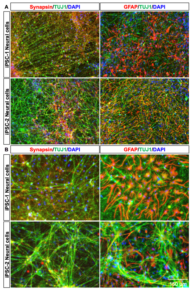
Immunofluorescent staining of neurons derived using the 2D method reveals the robust expression of neuronal markers such as TUJ1 (green) and synapsin (red), indicating well-differentiated neuronal networks. Additionally, GFAP (red), a marker of astrocytes, demonstrates the presence of glial cells alongside neurons, suggesting the development of a co-culture environment. Images from both female (iPSC-1) and male (iPSC-2) iPSC- derived neural cultures show consistent neuronal and glial marker expression, highlighting the reproducibility of the 2D method across different donor lines.
The 3D differentiation method yields neurons with distinct morphological characteristics and more intricate network formation compared to the 2D method. IF staining reveals pronounced TUJ1 (green) expression alongside synapsin (red), reflecting the formation of synaptic connections. GFAP (red) staining indicates the presence of astrocytes, and their three-dimensional arrangement mirrors the complexity of in vivo neural environments. Similar to the 2D method, the 3D protocol demonstrates consistent outcomes across female (iPSC-1) and male (iPSC-2) iPSC lines, reinforcing the reliability of this approach for generating advanced neural models.
These results highlight the capacity of both methods to generate neuron-astrocyte networks, with the 3D method offering enhanced structural complexity, suitable for modeling intricate CNS processes.